Dental Portable X-Ray Equipment:
With this project, in order to develop a portable digital dental x-ray system we aim to complete our scientific work and develop a new product which provides usage facilities with new specifications than existing X-ray-based imaging systems.
Product design continues in collaboration with Bogazici University, Institute of Biomedical BUMIL Laboratory and it is supported by San Tez.
The main objective of this project is to provide more economical operating costs for quite common dental x-ray filming, to reduce the dose of radiation in terms of making it more secure, to provide easier, low cost, fast and quality solution to the users and patients. With the help of to use CMOS-based direct digital sensor instead of film images, the images will be obtained with a minimum level of dosage to the user and patient. The images will be generated in DICOM standards and they will be shared instantly on computer environment. Image contrast enhancements and advanced measurement techniques will be developed with software and they can easily be modified with a simple user interface.
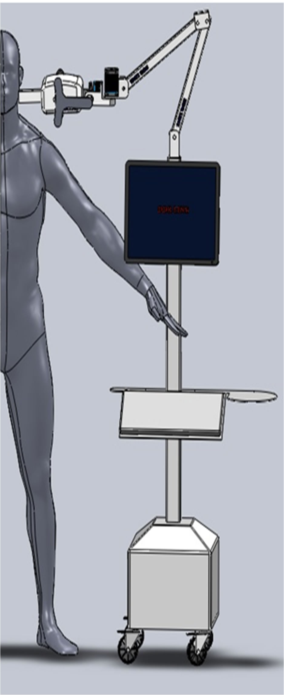
The most important feature of this system to be developed, it will operate without any cable connection with simple battery. Separate from the host device, the shooting part of the digital sensor, with a simple LCD, stand-alone without a plug and its being connected with a wireless communication system to the main console. With the help of this system it will be used in existing clinics to take shots from the patient’s location or several other locations with quickly carrying the system. The scattering radiation will be minimized with electronic and mechanic design extensions and according to that separate lead rooms will be unnecessary to use.Current Problems and Solution Suggestions:
Today, approximately 30% of the X-ray shots are taken for teeth. On the other hand, our country is currently quite behind of the overall technologic level about widespread used dental X-rays compared to the other X-ray-based applications like fluoroscopy, DSA and CT. General problems of the dentists listed as follows:
1. High / Unknown Dose: Replacing the tube only according to time, for the dental shots causes an uncertainty for the dose exposed at the shots. Tubes used with a single variable in daily use, leads to unnecessary dose propagation which is fixed to a one variable value only.
Solution: With the automatic sensor tube connection system, the radiation of the system will be controlled in real time. Sensor will report the needed radiation level to the tube, not only temporal, but also changing the voltage and current values which has a great importance in terms of contrast and image quality for the captured image and the optimum dose will be obtained.
2. Calibration: The current devices are used for many years without any test or control since their set up. In the process, the resulting change in performance of the X-ray tubes depending on factors, do not examined.
Solution: The dose of the tube system with automatic calibration system will be controlled continuously and an uncontrolled use of excessive radiation will not be allowed.
3. Image Processing: after obtaining the images, there aren’t any active processes to get a better quality and those images shown with very few details in monitors which are not suitable for clinical use.
Solution: The software modules for the needs of the clinics that are formed as a result of the meetings, which provides significant advantages in diagnosis. For example, root canal therapy in the image to automatically identify the channels and channel height measurement, examination of changeover time of the tooth structure is needed in orthodontics clinics and determining the placement of the implant for the implant treatments are few of these packages.
4. Scatter radiation: Most of the tubes used clinically pose serious risk about radiation emitting to its environment. Because of the cost and design deficiencies, the tubes which used for tooth applications expose the radiation to all around the room rather than exposing only to the tooth, and this poses a risk to doctors and patients.
Solution: Electronic design optimizing the dose of the radiation up to 80% decrease. Our device also blocks scattered radiation with mechanic radiation blockers.
5. Frequently Use: Increased temperature and problems at device operating cause patients to accumulate especially in busy clinics.
Solution: Users do not need to enter any variables to the system because of the fully automatic console. All images can be taken without moving the patients because of the portable system.6. Image Quality: Because of the use of poor quality sensors, unaligned tube and processing the images in low quality, cause serious anatomic losses in images.
Solution: Automatic sensor tube connection and taking the image in right angles using the robotic tube control system and transferring the image properly is our main goal for our devices which has DICOM medical imaging standards.
7. Fixed Indoor / Restricted Usage: Fixing the tubes to the walls, with reasons like high radiation rate and etc., restricted the use of the dental devices to limited places. This causes a clinical intensity and fragmentation in the treatment.
Solution: The portable device works fully independently from the electrical network and it is designed for the clinical usage. The treatments will be speed up with carrying the device to the patients.
8. Tube independent sensors: In current systems sensor and tube works and processes independently from each other and this causes difficulties for the image optimization and difficulty of use.
Solution: Automatic sensor tube connection transmits the needed parameters to tube from the sensor mechanism. System can optimize the radiation and can also calculate the needed parameters for the optimal image.9. Film Costs / Time problem: Bathing the films increase the amount of time for imaging to each film. In addition, the quality of bathing and user errors cause low quality images and it also increase the cost of consumables.
Solution: Fully digital system is compatible with the HIS and RIS structures of the hospital. With this way no problem occurs in image sharing and presenting. Internal high resolution monitors show all of the small details.Dual-Energy Direct Rontgen System
In this project we aim to decrease the radiation rate and increase the image quality to the top level with Direct Rontgen Systems which used frequently in general radiology for diagnostic. We plan to make high quality devices reachable to the users.
With the help of different powers of X-rays generated the contrast increases and high quality images formed. System hardware has advanced application features. For example, in lung images high sensitivity of the soft tissue may occur without ribs.
Dual Energy System with high contrast and image quality.
Save up to 50% of radiation
Fast rendering
High Energy Efficiency
50% more economical than imported systems
What is Direct Dual Energy X-Ray System?Dual energy X-ray technology is a technique that has emerged with the aim of bone densitometry measurements. The basis of the technique is to send two X-rays in different energies to obtain different contrasts. With establishing mathematical relationships between these values in the tissue we able to upgrade contrasts, or focus on desired piece of tissue.
In Densitometer systems, soft tissue contrast removed to reveal the bone mineral structure. In DSA, angio devices, images with and without contrast removed from each other and the great detail obtained for contrast images. These systems constitute a significant advantage in terms of doctors, especially for interventional treatments. With the development of new detector technologies, this process started to use more rapidly. The capacity of imaging, elevated with the help of new generation high frequency circuits which have reaction times in Milliseconds. These improvements about image processing have not been reflected yet in clinical practice which is technically possible.
With the help of new generation systems we are planning upgrade and apply this technology to direct imaging field.